
Staged L4–L5 Anterior to Psoas (ATP) Approach with Posterior Instrumentation
Patient Overview
54-year-old male with a BMI of 38.8 presenting with progressive low back pain, bilateral radiculopathy, and neurogenic claudication. The patient failed conservative management including physical therapy, medications, and epidural steroid injections.
Diagnosis
- L4–L5 degenerative spondylolisthesis
- Severe central canal stenosis
- Bilateral subarticular stenosis
Treatment Course
The patient initially underwent conservative management, including physical therapy and medications, without significant improvement. Symptoms progressed to neurogenic claudication and radiculopathy. MRI demonstrated severe central and foraminal stenosis at L4–L5. Bilateral L4–L5 transforaminal epidural steroid injections provided incomplete relief. Due to persistent and functionally limiting symptoms, the patient elected to proceed with surgical intervention.
Preoperative Imaging
Lateral Extension X-ray

Lateral Flexion X-ray
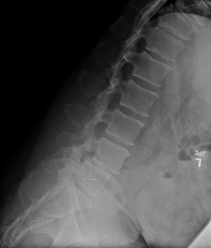
A/P X-ray

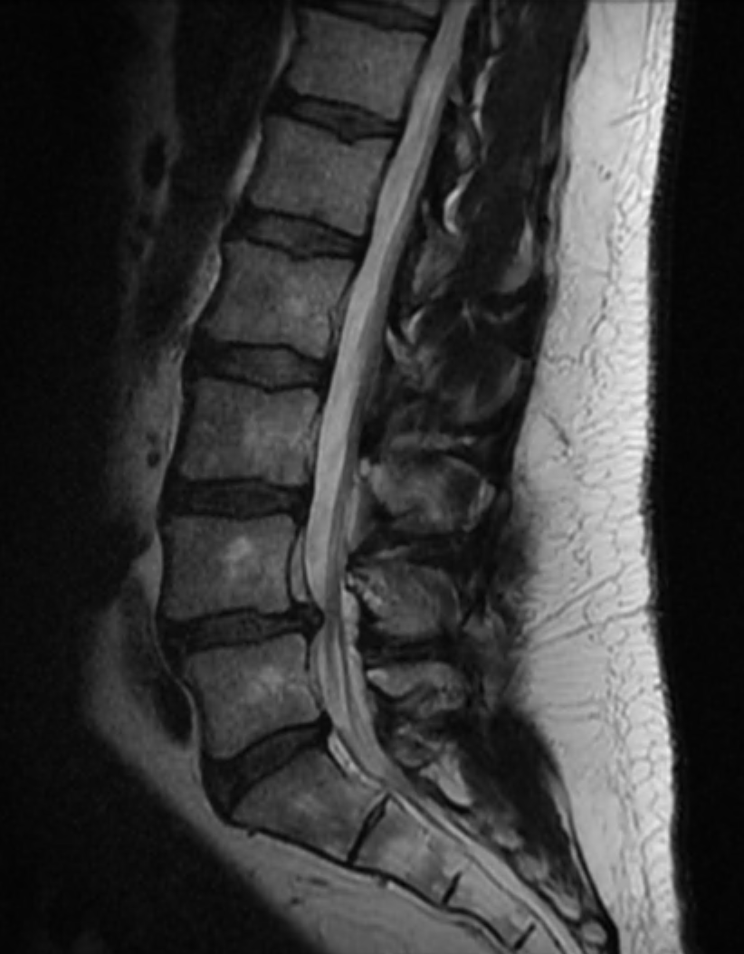
Sagittal MRI
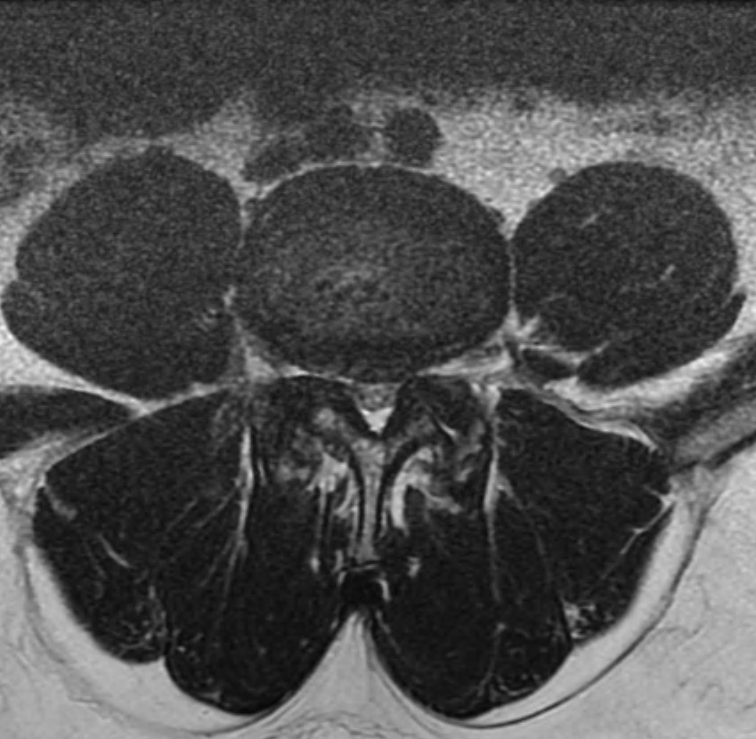
Axial MRI
Surgical Strategy
Stage 1 – ATP Approach
- L4–L5 anterior arthrodesis
- Interbody device placement
- Independent anterior plate fixation
- Bone marrow aspirate with allograft for fusion
Stage 2 – Posterior Instrumentation
- L4–L5 posterior arthrodesis
- Pedicle screw instrumentation
- Rod fixation
Orthofix Technologies Utilized
- Lateral lumbar interbody device (WaveForm™ L)
- Anterior plate system (Meridian™)
- Mariner™ pedicle screw system
- ATP instrumentation
- Lattus™ Retractor
- Bone marrow aspirate and allograft (OsteoStrand™ Plus)
ATP Instrumentation
Clinical Value
In this case, ATP instrumentation facilitated consistent exposure in a deep operative corridor in a high-BMI patient. The system provided stable retraction, improved visualization during disc preparation, and streamlined the overall workflow. This was particularly valuable in a technically demanding lateral anterior approach, allowing for efficient and precise implant placement.

Abilio Reis, MD
In patients with challenging anatomy and deep operative corridors, the ATP instrumentation has significantly improved my ability to achieve consistent exposure and maintain an efficient workflow. It allows me to focus on precise implant placement and achieve reliable surgical outcomes.
Physician Bio
Dr. Abilio Reis is a fellowship-trained spine surgeon at OrthoVirginia specializing in minimally invasive and robotic spine surgery. His practice focuses on advanced techniques to treat degenerative spine conditions and optimize patient outcomes.
Postoperative Outcome
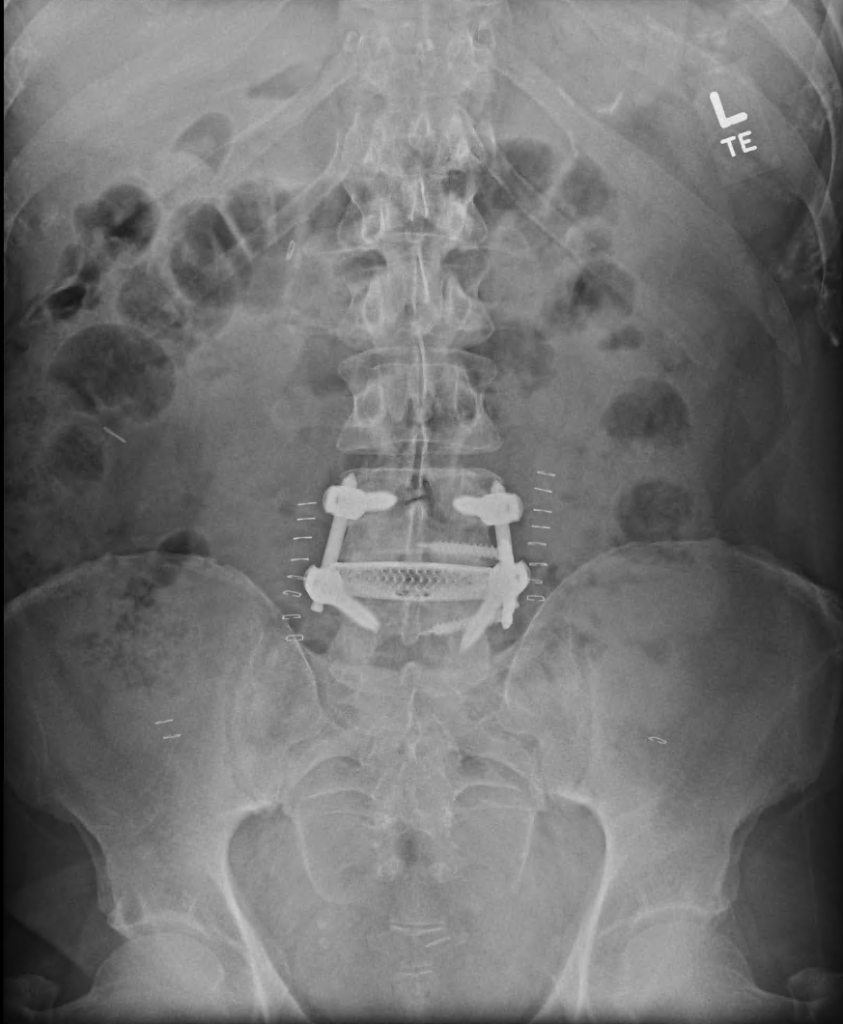
The procedure was completed without intraoperative complications. Estimated blood loss was approximately 50 ml per stage. Postoperative imaging demonstrated restoration of disc height, reduction of spondylolisthesis, and stable 360° construct with appropriate hardware positioning. The patient had a stable postoperative course.
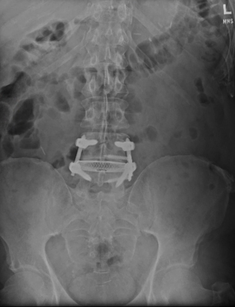
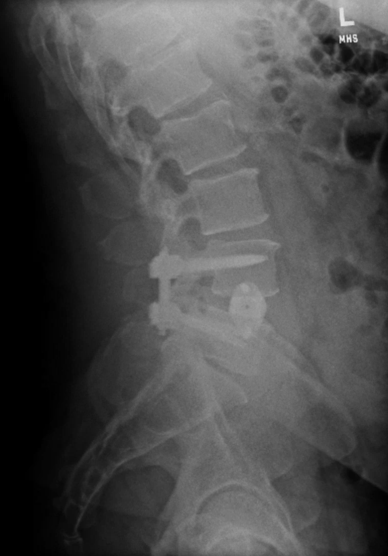
Intraoperative X-rays
A/P
Lateral

Postoperative X-rays
A/P
Lateral
The case report shows an individual’s response to treatment. The information contained in this case report is provided for informational and educational purposes. It is not intended to guarantee the response other people may have to treatment as individual results can and do vary. Proper surgical procedure is the responsibility of the medical professional. Each surgeon must evaluate the appropriateness of a technique based on his or her personal medical credentials and experience.